A cerebral infarction is a type of ischemic stroke that results from a blockage in the blood vessels supplying blood to the brain. This is the main cause of strokes, which caused the second largest number of deaths in 2013, after cancer. So far, patients had to have magnetic resonance imaging (MRI) and relied on doctors who examined their MRI images with subjective decisions in order to diagnose their symptoms. Thanks to this latest development, however, doctors will now be able to judge the riskiness of the disease backed by objective data.
The Korea Research Institute of Standards and Science (KRISS) along with 11 university hospitals, including Dongguk University Ilsan Hospital, formed the joint research team. The team focused on common symptoms of chronic ischemic strokes, which can be seen in patients suffering from cerebral infarctions. The researchers analyzed about 60,000 MRI images from a total of 2,699 patients who were admitted to the 11 hospitals over the past five years. According to 700 kinds of data points, including the age, weight and blood pressure of the patients, they categorized the amount of cerebral infarction, from one to 100, graded by age.
Dubbed as the “brain map of an ischemic stroke,” the accumulated data shows how much danger a patient faces with a table of images. If the patient faces a stronger danger of having an ischemic stroke, they will get a higher grade. Those who use the map can also check how healthy the patient's brain is by comparing MRI images with the brain map data, as the map provides images of brain vessels based on age groups.
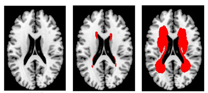
The above MRI images of the corona clinica in the brain are taken from patients who are aged between 65 and 74. The corona clinica is the central part of the cerebrum where motor and sensory nerves are located. The first grade image (left) shows barely any damage to the brain's blood vessels, while the images of the 50th and 100th percentiles (center, right) show partial or considerable damage to the blood vessels of the central part of the brain. The red color shows damaged blood vessels.
Patients can guess how much danger of an ischemic stroke they face by comparing their MRI images with the brain map.

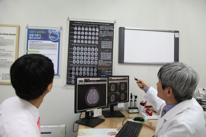
Dongguk University Ilsan Hospital Professor Kim Dong-eog
“With an ischemic stroke, there is a high possibility of death and disability. So prevention is absolutely important,” said Kim Dong-eog, professor at Dongguk University Ilsan Hospital. Kim was in charge of the research and who is chief of the Korea Brain MR Data Center. “Chronic ischemic strokes can be examined from MRIs of people who do not have any symptoms. It well-reflects how much danger the patient faces of an ischemic stroke. I hope many hospitals can use the brain map at work,” he added.
The researchers publicized their findings on the Korea Brain MR Data Center homepage (http://brainmr.com), allowing anyone to download the brain map and other related data. They will also make a panel for all hospitals so that their research results can be used for patients nationwide.
The results of the research were published in the December edition of Stroke, a U.S.-based international journal put out by the American Heart Association.
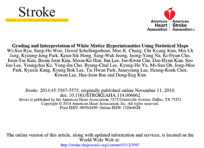
The brain ischemic stroke map is published in the December edition of Stroke.
By Yoon Sojung
Korea.net Staff Writer
Photos: Dongguk University Ilsan Hospital
arete@korea.kr
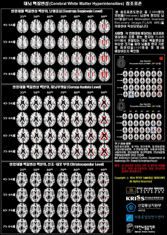
The KRISS and 11 participating university hospitals created a 'brain map of ischemic stroke' by making a standard medical chart based on the accumulated data collected from their patients. People can compare MRI images with the brain map.