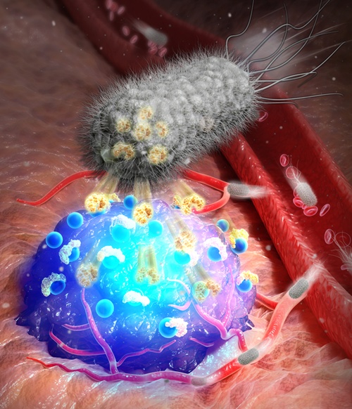
The newly developed bacterial imaging platform marks tumor locations with fluorescence by secreting streptavidin. (KIST)
By Koh Hyunjeong
A research team has developed a technology that uses bacteria to find where tumors are.
When combined with surgical robots or imaging assistance devices, this technology is expected to enhance real-time imaging accuracy and shorten surgery time.
Korea Institute of Science and Technology (KIST) on June 18 said the researchers developed a bacteria-based fluorescent imaging technology that enables during surgery real-time detection of cancerous areas by illuminating only cancerous tissue. The team comprised Suh SeungBeum of KIST's BioNics Research Center, Kim Sehoon of the school's Chemical and Biological Convergence Research Center, and Chungnam National University Hospital professor Lee Hyo-jin.
This method enables precise identification of cancerous areas by tracing fluorescent signals that persist for up to 72 hours.
Because cancer cells cause tissue necrosis, the principle is to take advantage of an oxygen-deficient environment by inserting a modified strain of the anaerobic bacterium salmonella into the body to seek cancerous areas.
Once the bacteria reach the cancerous cells, they send a signal to secrete streptavidin, which binds well to the vitamin biotin, and then a substance combining biotin and a contrast agent is injected into the body to locate the cells.
Suh said, "Engineering bacteria to autonomously seek cancer cells and send fluorescent signals allows the identification of the location and boundaries of the cancer in real time during surgery."
Precise removal of tumors during surgery is crucial. Preoperative imaging and ultrasound alone have proven insufficient to fully find the exact location and boundaries of tumors.
hjkoh@korea.kr
Most popular
- Military discharge sets stage for reunion of all 7 BTS members
- 'We are back!' BTS Festa heralds hyped return of K-pop phenom
- Presidents Lee, Trump discuss tariff deal in first phone talks
- President Lee leaves for G7 Summit in Canada on first int'l trip
- 'Maybe Happy Ending' wins 6 Tonys including Best Musical